The Scan After Morphology
Eager to catch a glimpse of your soon-to-arrive little one? A Growth Scan, typically performed in the third trimester, allows expectant parents the opportunity to see the remarkable progress their baby has made. This non-invasive ultrasound procedure is specifically designed to assess your baby’s size and overall health, offering invaluable insights and reassurance.
In this blog, we will discuss the fundamentals of a Growth Scan and provide insights into what you can expect during the procedure.
What Is Growth Scan?
As the name suggests, a Growth Scan evaluates your baby’s development during the final trimester of your pregnancy. It is generally conducted between weeks 22 and 40. This scan provides crucial information about your baby’s size and overall well-being, offering invaluable insights into their growth and development. The Growth Scan typically follows the Morphology Scan.
A third-trimester Growth Scan is often recommended for expectant parents who have a history of premature delivery, gestational diabetes, or other medical concerns that could impact the baby. It’s important to note that this scan is not a standalone diagnostic tool and may not identify all potential issues. Rather, it serves as an additional source of reassurance for parents and offers an opportunity to bond with their unborn child.
What to Expect During a Growth Scan?
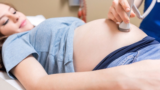
When you go for an ultrasound in Melbourne, you will be asked to relax and lie down on an examination table. A special gel will be applied to your abdomen and pelvic region. Subsequently, a small wand known as a transducer will be utilised to capture images of your baby, which will be displayed on the ultrasound screen.
For a Growth scan, your specialist will maneuver the transducer over the gel-covered area to view your baby from various angles, occasionally asking you to change positions or hold your breath to capture the best possible images. The scan is painless and safe for both you and your baby, generally taking between 30-45 minutes to complete.
During the Growth scan, your specialist will meticulously examine and measure various aspects of your baby’s growth and development, including:
During a dating scan, your baby’s ‘crown-rump length’ (CRL) is measured. This refers to your baby’s length from head to bottom. The CRL helps doctors examine the baby’s gestational age and probable due date.
1. Head Circumference:
To gauge the size of your baby’s head.
2. Abdominal Circumference:
To assess the size of your baby’s abdomen.
3. Femur Length:
To measure the length of your baby’s thigh bone.
4. Amniotic Fluid Levels:
To ensure adequate levels of amniotic fluid surrounding your baby.
5. Baby’s Position and Movements:
To monitor your baby’s activity in the womb.
6. Placental Position and Maturity:
To evaluate the placement and maturity of the placenta.
By comparing these measurements to growth charts and considering the number of weeks you are into your pregnancy, your specialist can determine how well your baby is progressing. They will also provide an estimated weight of your baby.
Crucial Body Parts Checked During the Scan
Most doctors recommend a Growth scan at around 36 weeks of pregnancy, a stage when teh majority of your baby’s essential body parts have developed. This allows the specialist to carry out a comprehensive examination, focusing on:
1. Brain:
To ensure that your baby’s brain is developing as expected.
2. Heart:
To assess the heart’s size, shape, and rhythm, ensuring proper blood circulation.
3. Diaphragm:
To verify the size and position of the diaphragm, essential for breathing.
4. Stomach:
To examine the stomach’s size and positioning for normal functioning.
5. Kidneys:
To check the kidneys’ size and location, which is crucial for waste removal.
6. Bladder:
To evaluate the size and location of the bladder, which is vital for waste elimination.
Furthermore, a Growth scan can also aid in detecting a rare congenital heart defect known as Ebstein’s anomaly, where the tricuspid valve forms abnormally. Early identification of this condition is crucial for appropriate medical intervention.
Keep in mind that each baby develops at an individual pace, and there can be considerable variations in growth from week to week. Your healthcare team will take these fluctuations into account when evaluating your baby’s development. During the scan, you’ll have the opportunity to view your baby on the screen and observe their movements within the womb—a reassuring and special moment for many parents.
After the scan, your referring doctor will review the findings with you, answering any questions you may have and advising on any necessary subsequent steps.
Conclusion
A Growth Scan in pregnancy is a safe and non-invasive method for monitoring your baby’s growth and overall well-being during the third trimester. These scans are generally available through your GP, but for those requiring serial Growth Scans, these can be scheduled with a specialist and may be supported by Medicare. It’s important to double-check your eligibility for Medicare-supported serial Growth Scans.
The third trimester is also a period when some parents may consider capturing 3D or 4D images of their baby. However, it’s essential to keep in mind that our primary focus during a Growth Scan is assessing the health of your baby.
Considering a Growth Scan? Book an appointment today.


